What Long COVID Does to Your Brain — And How We Knew Before Anyone Had a Name for It
Long COVID brain symptoms didn’t start making headlines until 2021. But in our neurofeedback clinic, we saw them a year earlier.
Not new clients. People who had finished their neurofeedback training. People whose brains had normalized — whose EEG biomarkers had stabilized, whose anxiety had quieted, whose focus had returned. They had done the work. They were done.
And then they weren’t.
One by one, they walked back through the door at our neurofeedback clinic in Colorado, describing their symptoms. Brain fog. Difficulty concentrating. Fatigue & dizziness. Sleep disruption. It was as if months of progress had been erased.
My wife and professional partner, Shari Johansson — a licensed professional counselor, board-certified neurofeedback specialist, and QEEG diplomate — pulled their brain maps. What she found stopped us both.
The most common findings were increased frontal and temporal alpha, with a slowed prominence and inflammatory high beta. Slowed alpha rhythms. Frontal dysregulation. Temporal lobe disruption. These weren’t subtle changes. The brain was telling a story, and the story was clear: something had happened to these people’s nervous systems since the last time we’d seen them.
The common thread? Every one of them had recently had COVID-19.
This was months before “Long COVID” had a name. Before the media started covering it. Before the medical establishment acknowledged it was real. We didn’t have a label for what we were seeing. But we had the data.
Why This Matters to You
If you’re reading this during International Long COVID Awareness Month in March 2026, you may be one of the millions of people who have experienced something similar — a cognitive shift that you can’t quite explain, that started after a COVID infection, and that hasn’t fully resolved.
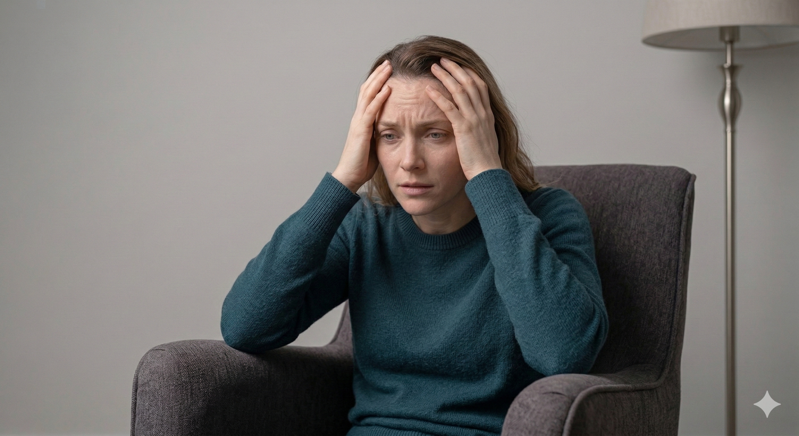
Maybe you describe it as “brain fog,” because that’s the closest word you have for the experience of reaching for a thought and finding it isn’t there. Maybe your reading comprehension has declined, or you’ve noticed your handwriting has gotten worse, or you can’t follow a conversation the way you used to. Maybe you’re just… slower.
You’re not imagining it. And you’re not alone.
According to research published in The Journal of Infectious Diseases, an estimated 3 to 5 million U.S. adults were experiencing activity-limiting symptoms of Long COVID as of late 2021 — and when accounting for underreporting, that number may have been as high as 9.7 million. The 2026 awareness campaign reports an estimated 400 million people worldwide have been affected at some point since 2019.
But here’s what most articles about Long COVID won’t tell you: there is a way to see what’s happening inside your brain. And once you can see it, there are tools — specific, evidence-based, non-invasive tools — that can help the brain recover. Long COVID brain dysfunction is not something you have to live with indefinitely.
That’s what this article is about. Not hype. Not miracle claims. Just what we’ve observed across five years of clinical work with Long COVID patients — backed by peer-reviewed research, personal experience, and hundreds of brain maps.
What COVID Does to the Brain: The Mechanisms
To understand why Long COVID produces cognitive symptoms, you need to understand how SARS-CoV-2 interacts with the nervous system.
The COVID virus is covered in spike proteins, which bind to ACE2 receptors throughout the body — including on neurons and the blood-brain barrier. Once bound, the virus can trigger a cascade of neurological damage through several pathways.
Neuroinflammation. When the virus activates the brain’s immune cells (microglia), it triggers a release of inflammatory cytokines — what clinicians call a “cytokine storm.” In an inflammatory state, overactive microglia produce reactive oxidative species (ROS) that damage neurons and disrupt normal brain function. This neuroinflammation is a major driver of the cognitive symptoms Long COVID patients describe.
Vascular damage and micro-clotting. Spike proteins damage the endothelial lining of blood vessels, including the tiny capillaries that supply the brain with oxygen. Micro-clotting in these vessels creates small areas of ischemia — reduced blood flow — that the brain experiences as slowed processing, difficulty with word retrieval, and impaired executive function.
Reduced cerebral blood flow. When the brain isn’t getting enough oxygen due to vascular damage and micro-clotting, overall brain metabolism drops. On an EEG, this appears as a slowing of brain rhythms — exactly the pattern we’ve observed in our clinic and that researchers worldwide have documented.
The spike protein persistence problem. Perhaps most concerning, spike proteins have been found in monocytes up to 15 months after COVID infection. This means the inflammatory process may continue long after the initial illness has resolved — which helps explain why symptoms worsen over time for many patients rather than improving.
There’s another mechanism worth understanding: COVID acts as a magnifier of existing neurological vulnerability. If you had a prior brain injury — whether from a concussion, Lyme disease, Epstein-Barr virus, or any other neurological insult — COVID doesn’t simply add to the damage. It amplifies it. In our clinical experience, this amplification appears to be exponential rather than additive. A person who was managing well despite an old concussion may find that post-COVID, those compensated symptoms come roaring back — and then some.
What the EEG Reveals: Seeing the Invisible
The challenge with Long COVID brain symptoms is that they’re invisible. Standard blood tests don’t show them. MRIs often come back normal. Patients are frequently told that nothing is wrong — or worse, that it’s “just stress.”
But the EEG tells a different story.
A quantitative EEG (QEEG) measures the electrical activity of the brain across 19 sites on the scalp, analyzing the power and distribution of different frequency bands — delta, theta, alpha, beta, and gamma. Each band corresponds to different brain functions: delta dominates during deep sleep, alpha reflects calm wakefulness and focus, beta drives active thinking and processing, and so on.
In healthy brains, these rhythms follow predictable patterns. In Long COVID brains, those patterns are disrupted in specific, measurable ways.
What the Research Shows
A 2021 systematic review published in the Journal of Clinical Medicine analyzed 17 studies on EEG changes in COVID-19 patients and found consistent abnormalities — particularly in the frontal regions of the brain. Patients showed slowing of brain rhythms, with increased slow-wave activity (delta and theta) and reduced healthy activity. A Cleveland Clinic study comparing 31 COVID-positive ICU patients to 38 age-matched controls found distinct EEG slowing — enhanced delta power with attenuated alpha and beta power. Strikingly, these changes were more severe in younger individuals (under age 70), which challenges the common assumption that older people are the most neurologically vulnerable to COVID.
A 2020 review of EEG data from 617 COVID-19 patients confirmed that frontal EEG abnormalities were common enough to be proposed as a biomarker for COVID-related encephalopathy.
What We’ve Observed in Our Clinic
Over the past several years, approximately 25% of our client load at Total Neuro Solutions has consisted of Long COVID patients. Many of these clients had not initially connected their cognitive decline to their COVID illness — particularly if their acute infection was mild. But the brain maps told the story.
The patterns we see most commonly in Long COVID QEEG assessments include:
- Generalized slowing of the EEG — the brain’s processing speed has literally decreased
- Left temporal slowing, often bilateral — which correlates with language processing difficulties, word-finding problems, and auditory processing challenges
- Slow edge of alpha indicating ischemia — reduced blood flow leaving a visible signature on the brain map
- Frontal slowing and encephalopathic patterns — impairing executive function, decision-making, and working memory
- Excess alpha and slowing of the alpha peak frequency — the brain’s “idle speed” has dropped
- Spindling excess beta (SEBs) — an inflammatory pattern often associated with anxiety, hypervigilance, and sleep disruption
These findings align precisely with what the peer-reviewed research describes. But they also tell us something the research hasn’t fully addressed yet: these patterns are treatable. The brain maps don’t just show damage — they show a roadmap for recovery.
When the Clinicians Became the Patients
I write about this subject not only as a neurofeedback practitioner and certified brain health coach, but as someone who lived it.
In late November 2021, a tile worker came into our home with visible signs of illness. My wife Shari was in the enclosed bathroom space where he was working. She contracted the Delta variant first. I became infected shortly after, caring for her at home.
Shari’s case was severe. She was hospitalized and admitted to the ICU on December 2nd. They told her she likely wouldn’t survive without a ventilator. She called me to say goodbye.
We refused the ventilator. We had studied the data and understood the risks. Instead, we advocated for her care — including stopping the high-sugar foods the hospital was providing while her medications had pushed her A1c into diabetic range. They switched her to eggs and protein. Within days, she was released. The nurse told me it was a miracle.
It wasn’t a miracle. It was informed decision-making under impossible pressure.
But the real work began at home. Shari could barely climb the stairs without severe shortness of breath and minutes of recovery after every exertion. She used an oxygen concentrator for months. In April 2022, when we took a weekend trip to Silverthorne, Colorado — elevation roughly 9,000 feet — her pulse ox dropped into the 80s. We had to buy canned oxygen to get her through.
Cognitively, everything was slowed. It was weeks before she could return to the office. When she did, she struggled to remember clients’ names. Processing EEGs — the very data she’s an expert in — took at least twice as long as it used to. Her background alpha had dropped. The overall power in her brain was very low.
She felt stupid. She was afraid she’d never recover. And she could see the evidence of her own decline every time she looked at her brain map.
My own case was less severe, but the cognitive effects were real. I noticed shortness of breath first, then congestion so thick I thought I’d suffocate at night. My pulse ox regularly dropped below 90. My fever stayed above 102 for days. By December 16th — roughly three weeks after onset — I was back on the ice playing goalie for my hockey team, because that’s who I am. The same push-through mentality that got me back to managing multi-million dollar electrical projects after six concussions.
But the shortness of breath on the ice felt like it was going to kill me. And in the weeks after returning to work, I noticed my handwriting was shaky. My typing speed had dropped dramatically. My speed of thought was slower. I’d always been able to listen to several conversations simultaneously — post-COVID, I struggled to focus on just the one in front of me. My spelling, something I’d always been excellent at, deteriorated noticeably.
I knew enough about brain function to recognize what was happening, even though I wasn’t yet working full-time in the neurofeedback field. These weren’t vague complaints. These were measurable, specific losses in cognitive function — the kind that show up on a QEEG as the exact patterns we now routinely identify in our Long COVID clients.
The Recovery Protocol: Energy First, Then Training
We were fortunate. As neurofeedback practitioners, we had access to the very tools we now recommend to our clients. And we learned something critical about the sequencing of recovery that shapes our clinical approach to this day.
You start with energy. Then you train.
When a brain is running on low power — when alpha amplitude has dropped, when overall EEG voltage is suppressed, when the person can barely climb stairs without losing their breath — you don’t begin with the intervention that demands cognitive effort. You begin with the one that delivers energy to the cells.
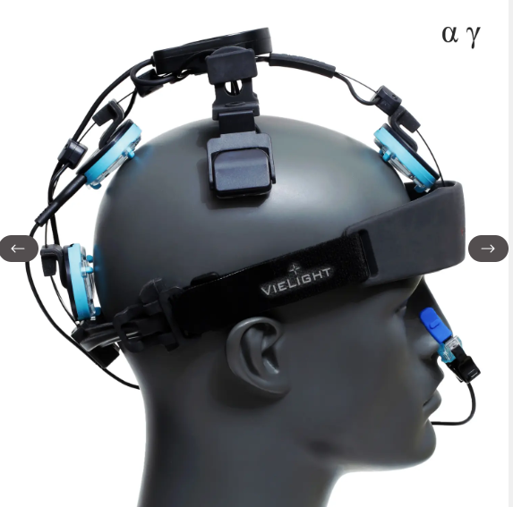
For us, that meant photobiomodulation (PBM) first. We had both the Neuronic transcranial helmet and Vielight intranasal devices. PBM is passive — you sit, you wear the device, and near-infrared light penetrates the skull to stimulate mitochondrial function at the cellular level. It increases ATP production, promotes nitric oxide release (which improves blood flow), and reduces neuroinflammation. The Vielight intranasal units also helped heal the damaged sinus cavity while increasing cerebral blood flow.
Only after PBM had begun restoring baseline energy to the brain did we layer in neurofeedback training and Neurofield stimulation (PEMF) specifically targeted at brain recovery.
This sequencing — energy restoration before active training — is a principle we now apply with every Long COVID client. The brain needs fuel before it can learn.
Why This Should Matter to You Right Now
March 2026 marks the 4th Annual International Long COVID Awareness Month. The 2026 campaign focuses on cardiovascular and multi-system impacts under the tagline “Every Heartbeat Counts.” More than 400 landmarks worldwide will light up teal on March 15th — Long COVID Awareness Day.
But awareness without action changes nothing. Long COVID brain recovery is possible — and it starts with seeing what’s actually happening.
If you’re experiencing persistent cognitive symptoms after a COVID infection — brain fog, difficulty concentrating, word-finding problems, memory issues, emotional reactivity, sleep disruption — a quantitative EEG can show you exactly what’s happening in your brain. Not guesswork. Not a vague diagnosis of “long COVID.” Specific, measurable patterns that point toward specific interventions.
And those interventions exist. Neurofeedback, photobiomodulation, neuromodulation, and targeted supplementation protocols are helping Long COVID patients recover cognitive function — sometimes after years of being told nothing could be done.
The brain is not a broken machine. It’s an adaptive system that responds to input, environment, stress, and care. When it gets the right support, it recovers.
We’ve seen it in our clients. We’ve seen it in each other. We’ve seen it in the data.
This article is the first in a series for International Long COVID Awareness Month 2026. Coming next: how photobiomodulation is helping restore brain function in Long COVID patients, and a deep dive into the combination therapies — from apheresis to hyperbaric oxygen to targeted supplements — that address Long COVID as the multi-system condition it truly is.
David Johansson is a neurofeedback practitioner, certified brain health coach, and the founder of TheBrainAndBody.com. He works alongside his wife Shari Johansson (MA, LPC, BCN, QEEG-D) at Total Neuro Solutions. Shari has presented nationally on the neurological impacts of Long COVID and the role of neurofeedback in recovery.
Affiliate Disclosure: Some links on this site are affiliate links, which means I may earn a small commission if you make a purchase through them. This comes at no additional cost to you. I only recommend products I have personally evaluated, used, or believe in based on my professional experience.
References
- Kopańska, M., Banaś-Żabczyk, A., Łagowska, A., Kuduk, B., & Szczygielski, J. (2021). Changes in EEG Recordings in COVID-19 Patients as a Basis for More Accurate QEEG Diagnostics and EEG Neurofeedback Therapy: A Systematic Review. Journal of Clinical Medicine, 10(6), 1300.
- Saab, C.Y., et al. (2022). SARS-CoV-2 Slows Brain Rhythms with more Severe Effects in Younger Individuals. Research Square (Preprint).
- Porges, S.W. (2020). The COVID-19 Pandemic is a paradoxical challenge to our nervous system: a Polyvagal Perspective. Clinical Neuropsychiatry, 17(2), 135-138.
- Tenforde, W.M., et al. (2023). Point Prevalence Estimates of Activity-Limiting Long-term Symptoms Among United States Adults ≥1 Month After Reported SARS-CoV-2 Infection. The Journal of Infectious Diseases, 227(7), 855-863.
- Antony, A.R. & Haneef, Z. (2020). Systematic review of EEG findings in 617 patients diagnosed with COVID-19. Seizure, 83, 234-241.
- International Long COVID Awareness (2026). 4th Annual Global Campaign. longcovidawareness.life